ASIA ELECTRONICS INDUSTRYYOUR WINDOW TO SMART MANUFACTURING
JEOL Reinforces System for High Throughput Workflow
JEOL Ltd. has introduced the JIB-PS500i Focused Ion Beam Scanning Electron Microscopy (FIB-SEM) system. Accordingly, as with the finer structure of advanced materials and advancing complexity of processes, evaluation techniques. These include morphological observation and elemental analysis require higher resolution and precision.
In the semiconductor industry as well as in the battery and materials fields, “higher precision” and “thinner sample” are necessary. Particularly, during the preparation of samples for transmission electron microscopes (TEM).
Most importantly, this product combines the FIB system that can process with high accuracy and the SEM of high resolution to satisfy these needs.
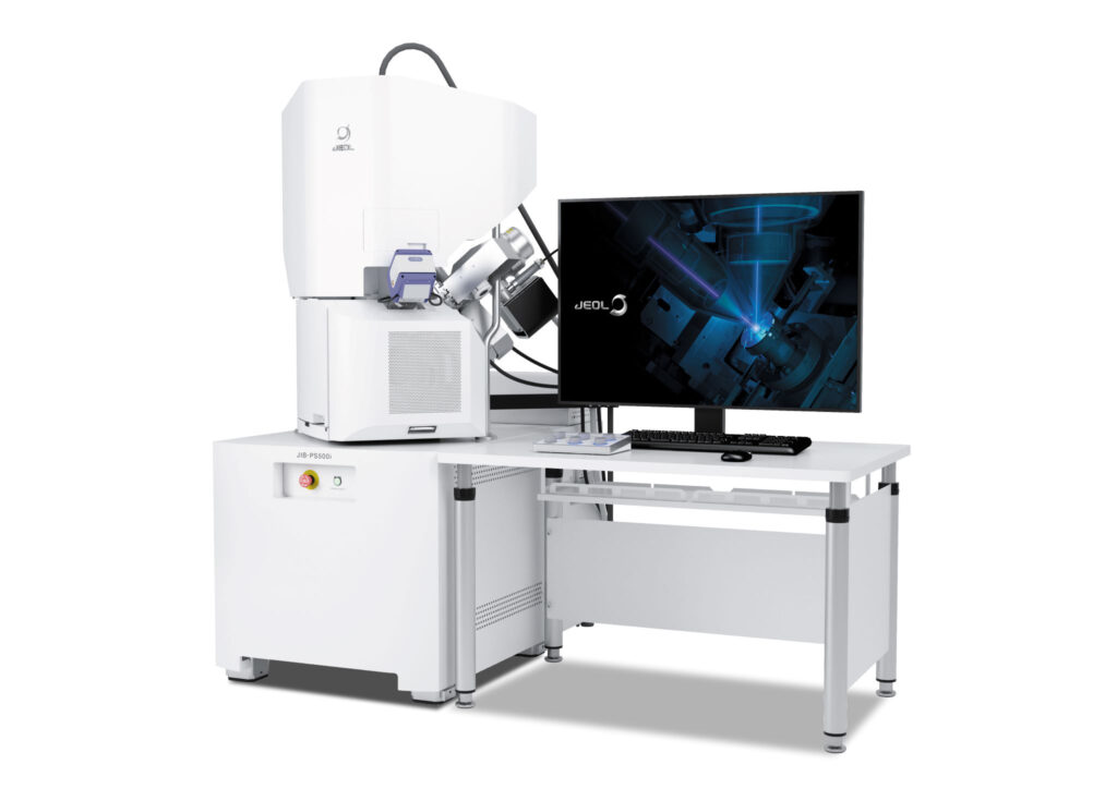
Main Features
The FIB column enables processing with a large-current Ga ion beam up to 100nA. The high-current processing is particularly effective in preparing cross section samples for large-area imaging and analysis. In addition, the FIB column sets to a shorter working distance. Along with a newly-developed power supply, it has led to greatly improved processing performance at a low accelerating voltage.
Moreover, it comes with the newly-developed super conical lens system is built into the SEM column, greatly improving the image resolution at a low accelerating voltage. Hence, this superb imaging is very useful to check the end-point milling status of lamella specimen using the SEM.
The JIB-PS500i adopts a large specimen chamber and a newly-developed specimen stage, increasing the stage movement range. Thus, accommodating a large specimen.
In addition, it is possible to use the newly-developed STEM detector with the stage tilt at 90 degrees, allows for a seamless transition from the TEM specimen preparation to STEM observation.
The operating GUI employs the SEM center, which has been well received in the JSM-IT800 series of high-resolution scanning electron microscopes. Hence, enabling full integration of EDS analysis. Furthermore, a double tilt cartridge and a dedicated TEM holder allows for more precise alignment while making specimen transfer between TEM and FIB easier.
- Share: